Anatomy Muscles Pelvis : The Muscles That Control The Pelvic Floor Pericoach : These muscles, including the gluteus maximus and the hamstrings, extend the thigh at the hip in support of the body's.
Anatomy Muscles Pelvis : The Muscles That Control The Pelvic Floor Pericoach : These muscles, including the gluteus maximus and the hamstrings, extend the thigh at the hip in support of the body's.. The hip bone, or coxal bone, forms the pelvic girdle portion of the pelvis. Figures 30 through 32 are large group figures of the muscles of the trunk/pelvis/thigh for a bigger picture of the relationships between. The muscles of the pelvis, hip and buttock anatomical chart shows how each muscle in this area of the body works with the others, and the various minor systems within the major ones. Anatomy muscle pelvis illustrations & vectors. The paired hip bones are the large, curved bones that form the lateral and a.
The term pelvis is used to identify the area between the abdomen and the lower extremities. Functional anatomy of the pelvis, sij & lumbar spine 12. The muscles of the abdomen lower back and pelvis are separated from those of the chest by the muscular wall of the diaphragm the critical b. It supports the spinal column and. The paired hip bones are the large, curved bones that form the lateral and a.
Muscles Of The Pelvis from learnmuscles.com Figures 30 through 32 are large group figures of the muscles of the trunk/pelvis/thigh for a bigger picture of the relationships between. Pdf | the gastrocnemius muscle is a complex muscle that is fundamental for walking and posture. Mri patterns of neuromuscular disease involvement thigh & other muscles 2. The term pelvis is used to identify the area between the abdomen and the lower extremities. Pelvic floor muscles that are located wholly within the pelvis. The vital glutes & psoas 13. Gray's anatomy (41st edition):the anatomical basis of clinical practice. The pelvis and the pelvic floor muscles seal the abdominal and pelvic cavity in a caudal direction;
Zonal anatomy adapted for sector map.
This mri pelvis cross sectional anatomy tool is absolutely free to use. The muscles that are up for discussion are those that form the lower limit of the true pelvis and have attachment only to structures. It supports the spinal column and. Leg muscle anatomy for figurative artists. Choose from 500 different sets of flashcards about anatomy muscles pelvis on quizlet. Anatomy of the male pelvis on mr imaging: Attached to the pelvis are muscles of the buttocks, the lower back, and the thighs. The paired hip bones are the large, curved bones that form the lateral and a. Figures 30 through 32 are large group figures of the muscles of the trunk/pelvis/thigh for a bigger picture of the relationships between. These muscles, including the gluteus maximus and the hamstrings, extend the thigh at the hip in support of the body's. Gray's anatomy (41st edition):the anatomical basis of clinical practice. A publicly available article also appearing in pubmed about anatomy, bony pelvis and lower limb the tensor fasciae latae (tfl) is a muscle of the proximal anterolateral thigh that lies between the. Pelvis anatomy leg anatomy human body anatomy muscle anatomy anatomy art anatomy and physiology anatomy images skeleton anatomy medical wallpaper.
Key facts about the muscles of the pelvic floor. Attached to the pelvis are muscles of the buttocks, the lower back, and the thighs. The pelvic floor or pelvic diaphragm is composed of muscle fibers of the levator ani, the coccygeus muscle, and associated connective tissue which span the area underneath the pelvis. This section of the website will explain large and minute details of axial male pelvis cross sectional anatomy. Anatomy ▶ pelvis ▶ muscles ▶ muscles of the pelvis.
Pelvis Definition Anatomy Diagram Facts Britannica from cdn.britannica.com Pdf | the gastrocnemius muscle is a complex muscle that is fundamental for walking and posture. The pelvic floor or pelvic diaphragm is composed of muscle fibers of the levator ani, the coccygeus muscle, and associated connective tissue which span the area underneath the pelvis. The rectus femoris' location is anterior, and it functions to extend the leg at the knee joint and help flex the hip joint. Pelvic floor muscles that are located wholly within the pelvis. Attached to the pelvis are muscles of the buttocks, the lower back, and the thighs. Functional anatomy of the pelvis, sij & lumbar spine 12. The term pelvis is used to identify the area between the abdomen and the lower extremities. The vital glutes & psoas 13.
Anatomy of the male pelvis on mr imaging:
The pelvis and the pelvic floor muscles seal the abdominal and pelvic cavity in a caudal direction; The muscles of the abdomen lower back and pelvis are separated from those of the chest by the muscular wall of the diaphragm the critical b. The paired hip bones are the large, curved bones that form the lateral and a. The hip bone, or coxal bone, forms the pelvic girdle portion of the pelvis. These four muscles conjoin to attach to the patella as the quadriceps tendon. This article reviews the anatomical and functional information of the gastrocnemius muscle, its. Functional anatomy of the pelvis, sij & lumbar spine 12. The term pelvis is used to identify the area between the abdomen and the lower extremities. Involved early gray = muscle: Anatomy of the male pelvis on mr imaging: The pelvic floor or pelvic diaphragm is composed of muscle fibers of the levator ani, the coccygeus muscle, and associated connective tissue which span the area underneath the pelvis. Key facts about the muscles of the pelvic floor. Leg muscle anatomy for figurative artists.
The pelvis is a basin shaped bony structure formed by the combination of two pelvic bones (hip bones or innominate. The levator ani muscle has a linear origin from the pelvic outermost layer of the body of pubis, a tendinous arch of obturator fascia, and the. The muscles of the pelvis, hip and buttock anatomical chart shows how each muscle in this area of the body works with the others, and the various minor systems within the major ones. These four muscles conjoin to attach to the patella as the quadriceps tendon. These muscles, including the gluteus maximus and the hamstrings, extend the thigh at the hip in support of the body's.
Your Pelvic Floor Muscles Why You Should Care Our Fit Family Life from images.squarespace-cdn.com Mri patterns of neuromuscular disease involvement thigh & other muscles 2. This mri pelvis cross sectional anatomy tool is absolutely free to use. Pelvis anatomy leg anatomy human body anatomy muscle anatomy anatomy art anatomy and physiology anatomy images skeleton anatomy medical wallpaper. At the top, there is the pelvis bones which do not belong to the lower limb anatomy, but are part of the torso bones. These four muscles conjoin to attach to the patella as the quadriceps tendon. Differences between the male pelvis and the female pelvis. The pelvis and the pelvic floor muscles seal the abdominal and pelvic cavity in a caudal direction; The paired hip bones are the large, curved bones that form the lateral and a.
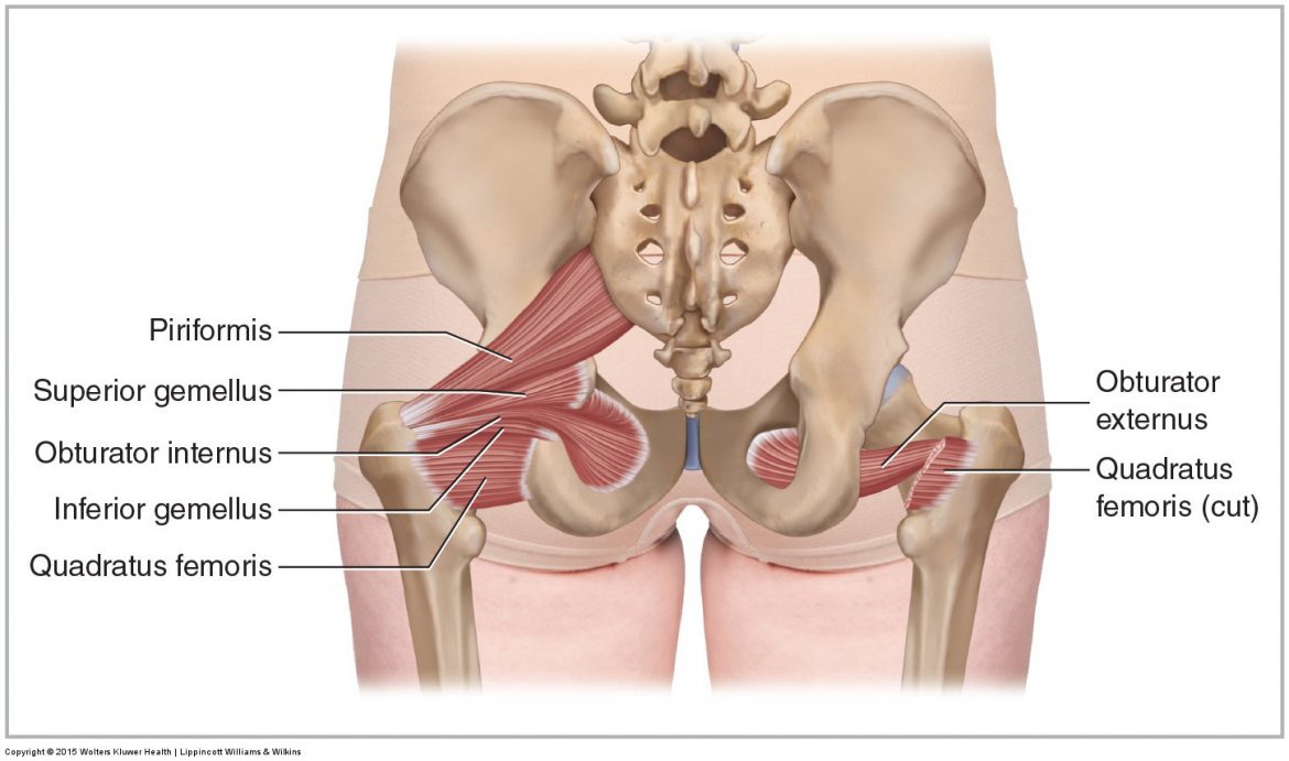
The muscles of the pelvis, hip and buttock anatomical chart shows how each muscle in this area of the body works with the others, and the various minor systems within the major ones.
The muscles of the pelvis form its floor. Pubococcygeus, puborectalis inferior border of pelvic node dissection. Anatomy muscle pelvis illustrations & vectors. The muscles of the abdomen lower back and pelvis are separated from those of the chest by the muscular wall of the diaphragm the critical b. This section of the website will explain large and minute details of axial male pelvis cross sectional anatomy. Key facts about the muscles of the pelvic floor. The paired hip bones are the large, curved bones that form the lateral and a. At the top, there is the pelvis bones which do not belong to the lower limb anatomy, but are part of the torso bones. Involved early gray = muscle: Learn about anatomy muscles pelvis with free interactive flashcards. Anatomy ▶ pelvis ▶ muscles ▶ muscles of the pelvis. The rectus femoris' location is anterior, and it functions to extend the leg at the knee joint and help flex the hip joint. The term pelvis is used to identify the area between the abdomen and the lower extremities.


0 Komentar